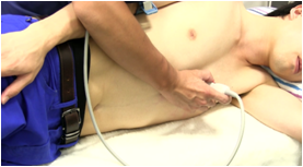
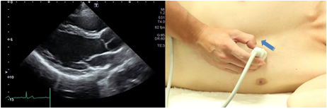
Left ventricular longitudinal view
 |
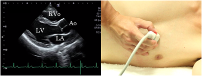 |
Parasternal views usually are obtained with the patient in the lateral decubitus position to avoid the effects of air contained in the lungs. To obtain a left ventricular longitudinal view, place the probe on the parasternal area at the 3rd or 4th intercostal space to visualize the left ventricle at the maximum inner diameter and the interventricular septum and the anterior wall of the aorta at almost the same height. This view is suitable for observation of the left ventricle, the right ventricle, the mitral valve, the aortic valve, and the proximal part from the Valsalva sinus to the ascending aorta.
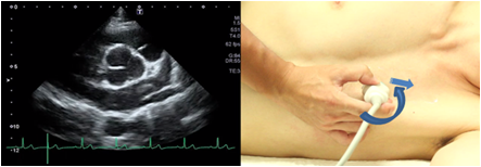
From the anterior commissure to the posterior commissure
 |
 |
The anterior commissure is visualized when tilted the probe slightly outward from the mitral valve mid portion so that the ultrasound beam is directed to outer side. At this time, adjust the probe so that the anterior papillary muscles, chordae tendineae, and the mitral valve are visualized in succession. The posterior commissure is visualized when tilted the probe slightly inner ward from the mitral valve mid portion so that the ultrasound beam is directed to inner side. These views are suitable to observe the site of a prolapsed mitral valve or a lesion of mitral valve stenosis and subvalvular apparatus.
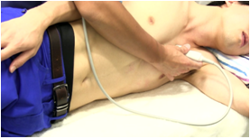
High intercostal approach
 |
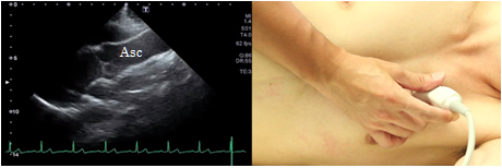 |
In the high intercostal approach, place the probe along the parasternal long axis at one or two intercostal spaces above the depiction position of the longitudinal parasternal view of the left ventricle and try to visualize it to the peripheral side of the ascending aorta. This view is suitable to observe lesions in the ascending aorta.
Longitudinal view of the right ventricular inflow tract
 |
 |
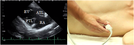 |
To obtain the longitudinal view of the right ventricular inflow tract, rotate the probe slightly in a clockwise direction from the longitudinal parasternal view of the left ventricle and tilt it so that the ultrasound beam is directed slightly downwardly. The anterior cusp of the tricuspid valve is visualized in front and the posterior cusp is visualized at the back. This view is suitable for observation of the tricuspid valve, right ventricle, and the right atrium and to estimate the right ventricular systolic pressure from tricuspid regurgitation.
Longitudinal view of the right ventricular outflow tract
 |
|
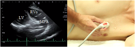 |
To obtain a longitudinal view of the right ventricular outflow tract, rotate the probe slightly in a counterclockwise direction from the position showing the aortic level of the short axis view of the left ventricle and tilt it so that the ultrasound beam is directed slightly outwardly. This view is suitable for the determination of wall motion abnormalities of the right ventricular outflow tract, stenosis of the right ventricular outflow tract, and pulmonary valve stenosis.