Apical longitudinal view
 |
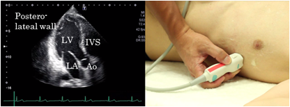 |
To obtain the apical longitudinal view, place the probe at the apex located at the apical impulse. This view is suitable for evaluation of the wall motion from the left ventricle anterior septum, apex and left ventricular posterior wall. It is also suitable for recording of the left ventricular inflow and left ventricular ejection velocity using the Doppler method and evaluation of aortic stenosis.
Apical 4 chamber view
 |
The apical 4 chamber view is found by placing the probe at the apex located at the apical impulse. The left chambers are seen on the right of the screen and the right chambers on the left of the screen. Fix the image when the 4 chambers look the largest. The important point is to visualize the left ventricular apex directly under the probe on the screen. This view is suitable for evaluation of 4 chamber balance and wall motion abnormalities and abnormalities of the attachment site of atrioventricular valves. It is also suitable for the measurement of left ventricular volume, left ventricular ejection rate, and for recording of pulmonary venous blood flow patterns. |
Apical 2 chamber view
 |
 |
The apical 2 chamber view can be obtained by the 90 degrees counterclockwise rotation of the probe from the position of visualizing the apical 4 chamber view. The proper view does not include the right chamber or the anterior papillary muscle. This view is suitable for evaluation of wall motion abnormalities in the anterior and inferior walls of the left ventricle and measurement of left ventricular volume and left ventricular ejection rate.